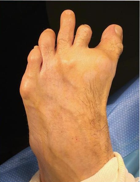
Complex bunion and hammertoe reconstruction
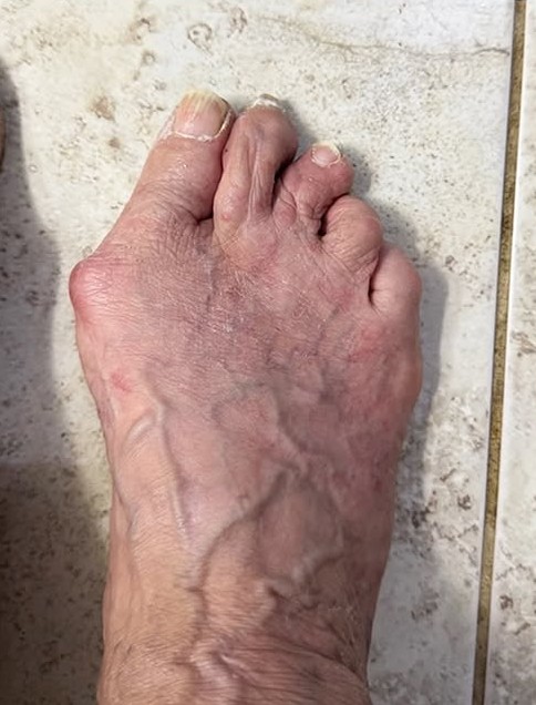
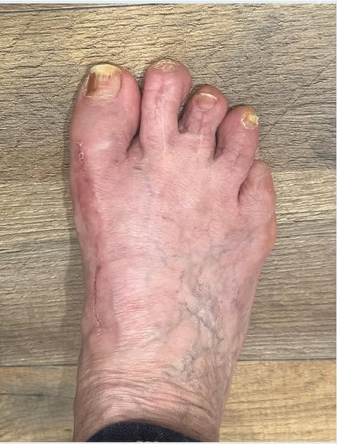
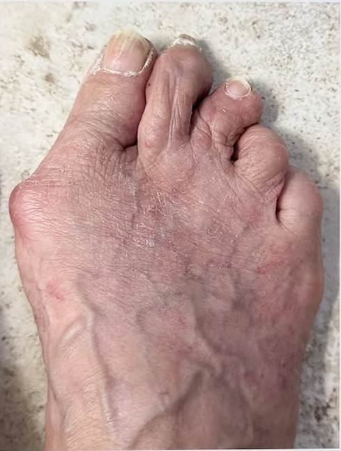
The before image shows an advanced bunion, medically known as hallux valgus, with the great toe drifting toward the smaller toes. Several lesser toes are also contracted and overlapping, including a pronounced crossover second toe. The after image shows the healed result following complex forefoot reconstruction: the great toe is straighter, the bunion prominence is reduced, and the lesser toes are in a more functional position.
Fifth-toe hammertoe correction
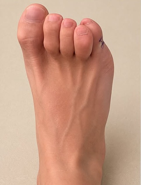
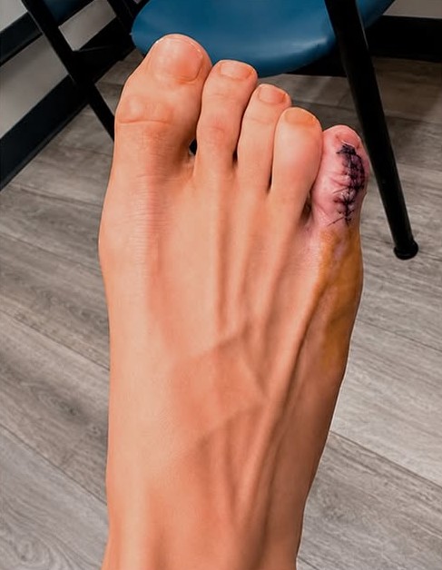
The fifth toe was rotated and curled inward toward the fourth toe, a deformity often called a curly toe, adductovarus toe, or fifth-toe hammertoe. The postoperative image shows the toe in a straighter position. The sutures indicate that the photograph was taken during the early healing period rather than after final recovery.
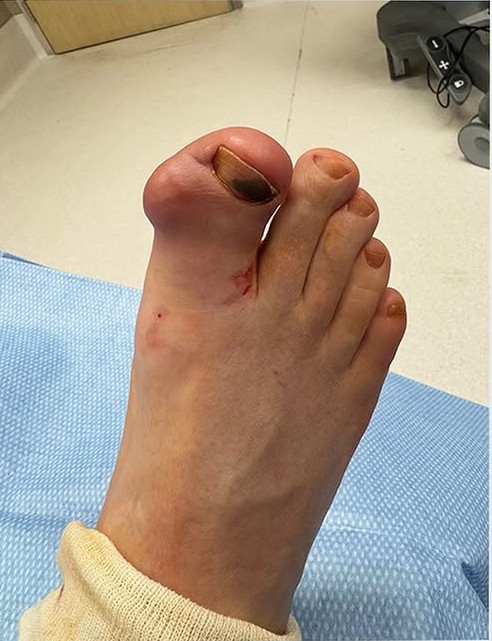
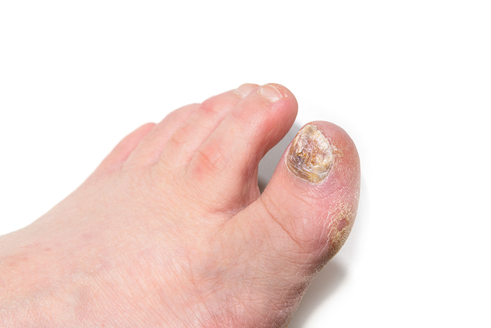
Treatment of a discolored fungal toenail
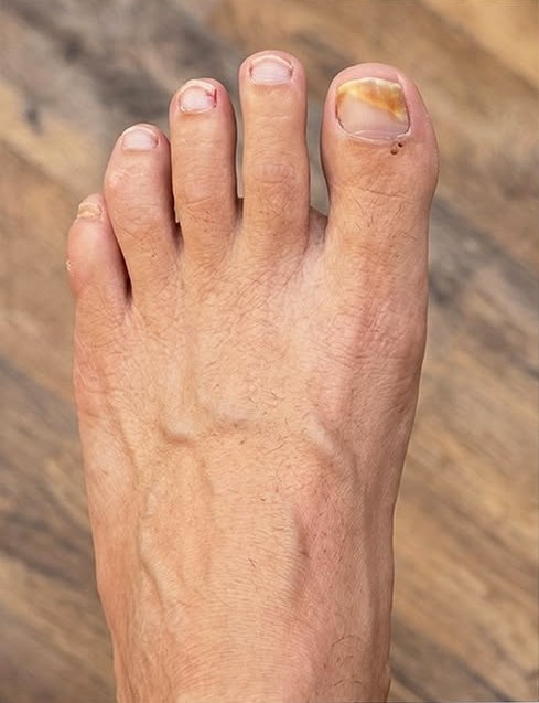
The before image shows yellow-brown discoloration involving the great toenail, an appearance commonly associated with toenail fungus. The after image shows clearer, healthier-looking nail growth as the damaged portion has grown out. Because nail trauma can resemble a fungal infection, the medical record should confirm the diagnosis and whether this result followed Lunula Laser, medication, debridement, or combination therapy.
Immediate postoperative forefoot reconstruction
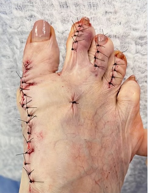
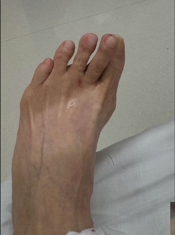
This appears to be the same complex case shown in Case 1, photographed immediately after surgery. The procedure addressed the severe bunion, crossover toe, and multiple hammertoe deformities. The incisions and sutures show where the bones, joints, tendons, or supporting tissues were surgically realigned. Case 1 appears to show the later, healed result.
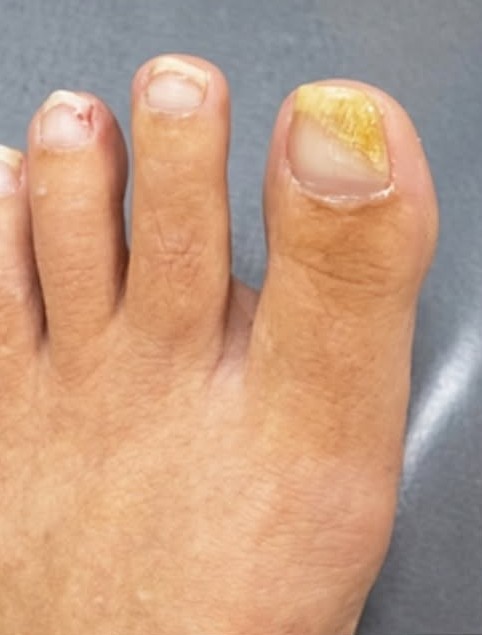
Improvement in great-toenail discoloration
The before image shows localized yellow-brown discoloration of the great toenail and irritation near the nail fold. The follow-up image shows reduced discoloration and healthier surrounding skin. The doctor should confirm whether this was fungal nail treatment, recovery from nail trauma, or treatment of another nail condition before the caption identifies a specific diagnosis.
Immediate postoperative forefoot reconstruction
This appears to be the same complex case shown in Case 1, photographed immediately after surgery. The procedure addressed the severe bunion, crossover toe, and multiple hammertoe deformities. The incisions and sutures show where the bones, joints, tendons, or supporting tissues were surgically realigned. Case 1 appears to show the later, healed result.
Hammertoe fusion with internal implants
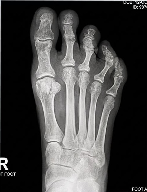
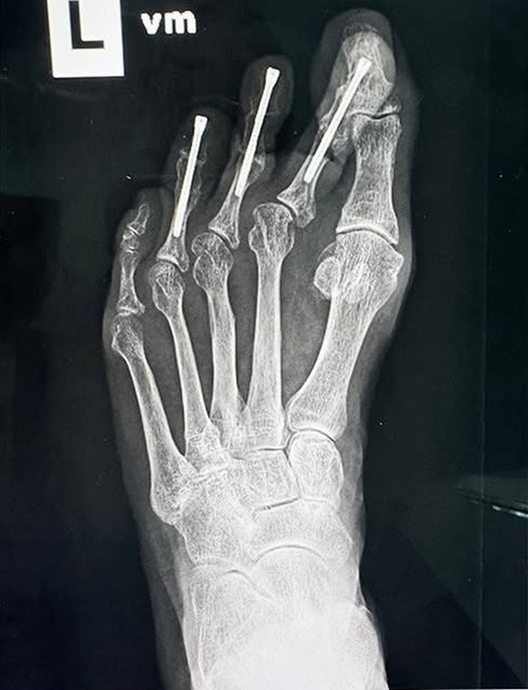
The preoperative X-ray shows rigid hammertoe deformities involving the second, third, and fourth toes. In the postoperative X-ray, the toes have been straightened and small internal screws span the corrected toe joints. These implants hold the toes in position while the joints fuse and heal—a procedure known as hammertoe arthrodesis.
Revision of previously operated hammertoes
The before X-ray shows existing implants in several lesser toes together with persistent or recurrent toe misalignment. The postoperative X-ray shows revised fixation, improved alignment, and a temporary pin supporting one of the corrected toes. This appears to be revision hammertoe surgery, but the doctor should confirm the precise reason for revision and the procedures performed.
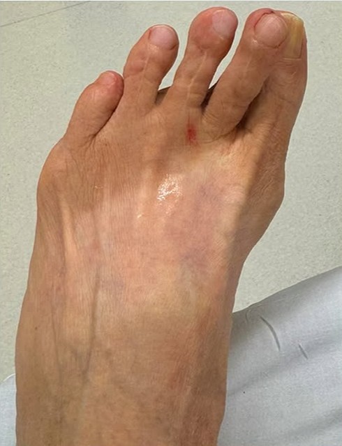
Immediate postoperative bunion correction
The before image shows a prominent bunion with the great toe deviating toward the second toe. The postoperative image shows the great toe placed in a straighter position, with sutures along the inside of the big-toe joint. This is an early postoperative view, so swelling and bruising are expected parts of the healing process. The doctor should specify whether this was Lapiplasty, an osteotomy, or another bunion-correction technique.
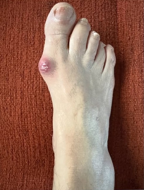
Healed Lapiplasty bunion correction
The before image shows hallux valgus with a painful-looking, inflamed prominence at the base of the great toe. The healed image shows a straighter great toe, a substantially reduced bunion prominence, and a well-healed surgical area. The clinic’s corresponding public post identifies this result with Lapiplasty bunion surgery.
Complex bunion and multiple-hammertoe reconstruction
The before image shows a complex forefoot deformity involving the great toe and several contracted, elevated, or displaced lesser toes. The immediate postoperative image shows correction of the bunion and multiple lesser-toe deformities. Temporary pins maintain the corrected toe positions during early healing, while the sutures mark the different areas addressed during surgery.
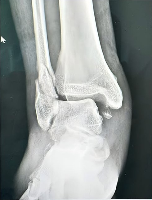
Surgical repair of an ankle fracture
The first X-ray shows a displaced ankle fracture, including a break of the distal fibula on the outside of the ankle. The postoperative X-ray shows the fractured bone restored to improved alignment and stabilized with a plate and screws. This procedure is called open reduction and internal fixation, or ORIF. The hardware holds the fracture securely while the bone heals and helps restore ankle-joint alignment.
Lesser-toe and crossover-toe correction
The before image shows drifting and deformity of the lesser toes. The postoperative image shows the toes in improved alignment, with surgical incisions over the corrected areas and a temporary pin supporting one toe during healing. The exact combination of hammertoe correction, tendon balancing, plantar-plate repair, or metatarsal realignment should be confirmed from the operative record.
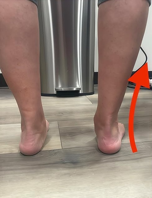
Improvement in rearfoot and heel alignment
Viewed from behind, the before image shows the heel positioned at an abnormal angle relative to the lower leg, an appearance that may be associated with pronation or flatfoot deformity. The after image shows the heel in a more vertical position beneath the leg, creating improved rearfoot alignment. The photographs alone do not establish whether this change resulted from custom orthotics, bracing, physical treatment, or reconstructive surgery, so the doctor should confirm the treatment.
Mosaic plantar-wart treatment
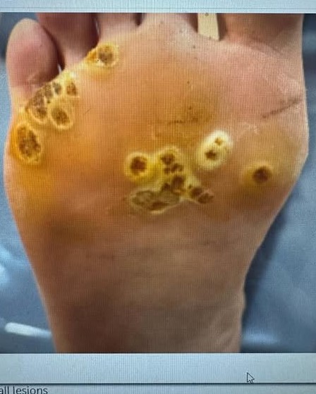
The before image shows numerous thick, rough lesions on weight-bearing areas of the sole, including clusters consistent with mosaic plantar warts. The after image shows a substantial reduction in the number, thickness, and size of the lesions. Several remaining areas are outlined, suggesting that the photograph may document progress during treatment rather than final complete clearance. The clinic’s public materials indicate that it uses Swift microwave therapy for plantar warts.
Plantar-wart treatment — currently missing from the page
The unpublished Case 15 images show a large, thickened plantar lesion before treatment and a much flatter, less prominent area afterward. The appearance is consistent with a treated plantar wart surrounded by pressure-related callus. The doctor should confirm whether this result followed Swift therapy and whether the after image represents completed treatment or an interim response.